Ultrasound scanner
An ultrasound machine with probes for examining the abdomen, chest, heart (echocardiography) and superficial tissues.
What an ultrasound machine consists of
Click or hover over a part. The descriptions appear one after another.
- 3Probes
A set of probes: convex for the abdomen, linear for superficial structures, phased array for the heart. They are chosen to suit the size of the patient, from a small cat to a large dog.
- 4Console with dials
Adjustment of depth, gain and image focus without looking away from the monitor.
- 5Central unit
A computer that converts the signal from the probe into an image and stores the archive of examinations.
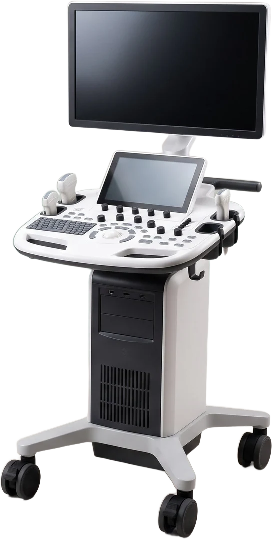
- 1Monitor
A real-time image: 2D mode, M-mode and Doppler. On it the veterinarian assesses the organs and measures their dimensions.
- 2Touch panel
Selection of the probe, examination settings and imaging mode; this is also where images and measurements are saved to the patient's record.
- 6Wheeled base
The machine moves between the ultrasound room, the operating theatre and the in-patient hospital, so the examination can be done at the patient's side.
Illustrative diagram of an ultrasound machine. The arrangement of parts corresponds to our set-up, but the appearance of the device may differ.
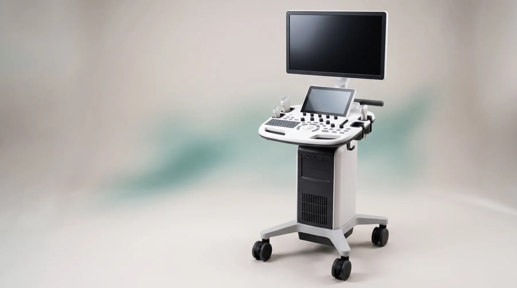 VINNO E35-style ultrasound machine
What it is and how it works
Ultrasonography images soft organs in real time using sound waves, without radiation. The set of probes makes it possible to examine both small cats and large dogs: the abdomen, bladder, kidneys, liver, intestines, lymph nodes and, with the phased array probe, the heart in 2D mode, M-mode and Doppler.
What it offers
The examination is painless and in most cases takes place without sedation, with the owner present. We discuss the result straight away, on the screen.
Ultrasound answers questions that X-ray cannot: the structure of an organ, the presence of fluid, blood flow, intestinal motility, and the size and shape of lesions. It also makes targeted biopsy under image guidance possible.