
Digital X-ray
A digital X-ray unit — the image is ready in a dozen or so seconds, with no film processing, and can be processed digitally and sent to the referring veterinarian.
What an X-ray suite consists of
Click or hover over a part. The descriptions appear one after another.
- 1X-ray tube with collimator
The radiation source. The collimator narrows the beam to the area being examined, limiting the dose to the patient and staff.
- 2Column
The tube stand: height and angle can be adjusted without moving the animal.
- 3Table with digital detector
A detector under the tabletop sends the image to the monitor within seconds, with no films or processing.
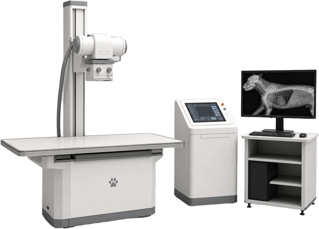
- 5Reporting workstation
Assessment of the image, magnification, measurement of angles and lengths (for example for planning TPLO) and archiving in the patient's record.
- 4Generator console
Selection of exposure settings for the species, body weight and the area being examined.
Illustrative diagram of a digital radiography system. The arrangement of parts corresponds to our X-ray suite, but the appearance of the devices may differ.
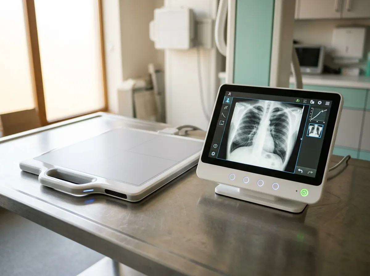 X-ray suite
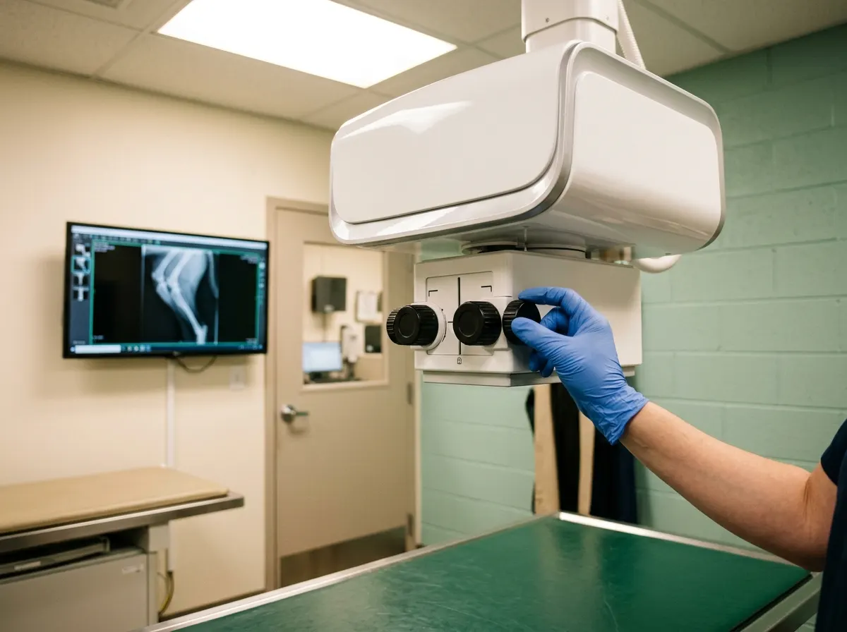 Assessment of the digital image
What it is and how it works
Digital radiography (DR) replaces conventional films with a detector that sends the image directly to the monitor. This allows the quality of the image to be assessed immediately, a view to be repeated without additional waiting, magnification, measurement of angles and lengths (for example when planning TPLO) and archiving in the patient's record.
What it offers
A shorter examination time means less stress for the animal and less frequent need for sedation. The owner receives the images in digital form and can take them away or pass them to another veterinarian.
High resolution and image processing reveal subtle changes in bone, fluid and lung. Measurements on the digital image are the basis for planning orthopaedic procedures.
Examinations and procedures where we use this
Our X-ray suite is safe
For animal diagnostics the practice uses, among other things, devices that produce ionising radiation: X-ray imaging machines. The X-ray suite in which this device is used was put into operation on the basis of a permit issued by the President of the National Atomic Energy Agency. The practice monitors the exposure of staff and bystanders using individual and environmental dosimetry carried out by an accredited laboratory. The practice's staff have been trained in radiation protection by a qualified radiation protection officer from the company RADIOVET – radiovet.pl.
Date: 01.09.2026