Specialty

Diagnostic imaging

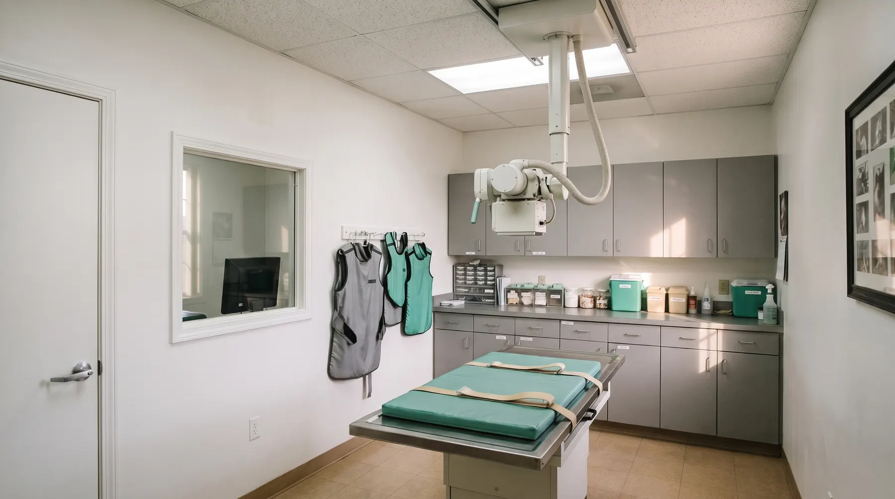
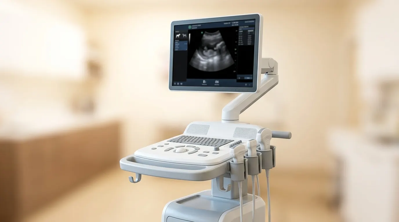
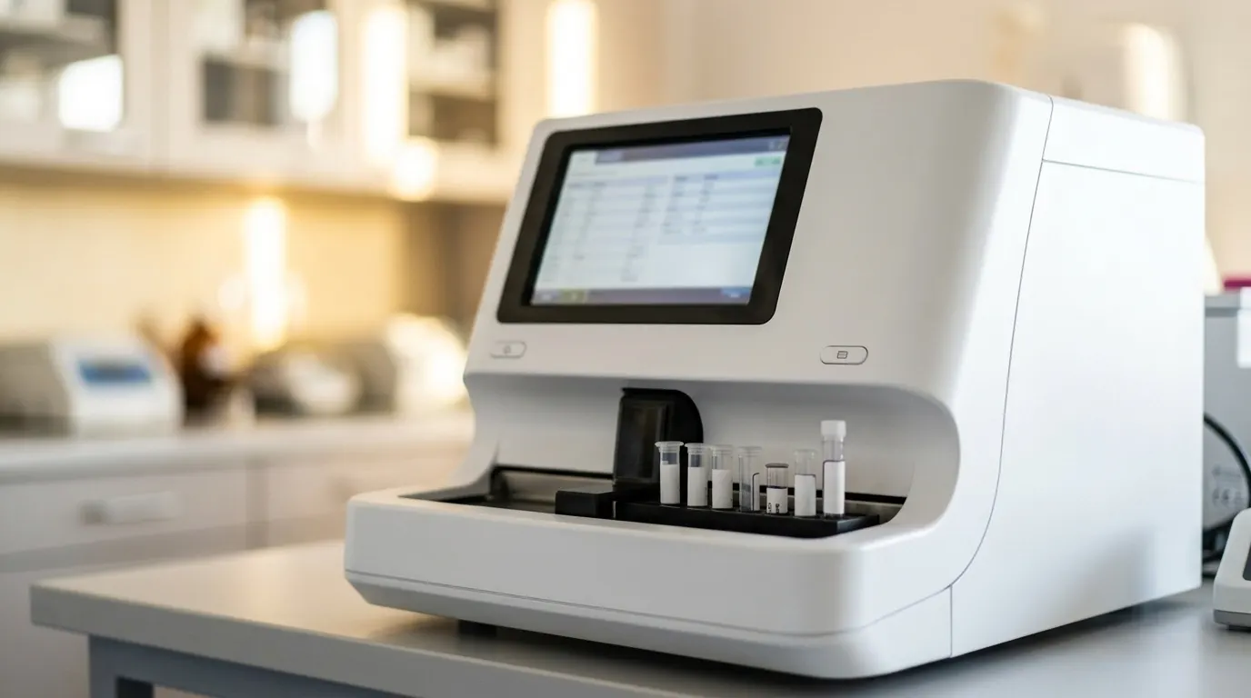
On-site digital X-ray and ultrasonography, reported in the patient’s clinical context — not “in passing”.

6 examinations and procedures5 items of equipment
Scope of this specialty

Examinations, procedures and services

We manage every case individually — the scope, indications and course described above are indicative; the exact diagnostic and treatment plan is agreed during the consultation.

Imaging · examination

Digital X-ray — radiography

Digital X-ray examination of a dog or cat, with the report given on site. Bones and joints, chest, abdomen, looking for foreign bodies — high-resolution images available immediately.

DogCat
Imaging · examination

Chest X-ray

Examination of the lungs, heart, trachea and oesophagus in a dog or cat with a cough, breathing difficulty or cancer. Three views, assessment on site, supplemented with a heart ultrasound if needed.

DogCat
Imaging · examination

X-ray for joint dysplasia

X-rays of the hip and elbow joints under sedation, in the positioning required to assess dysplasia — for breeding purposes or to investigate lameness in a young dog.

Dog
Imaging · examination

Abdominal ultrasound

A painless examination of the abdominal organs of a dog or cat — liver, kidneys, bladder, intestines, spleen, prostate and uterus — with the report given on site. The basis for investigating vomiting, diarrhoea, urinary problems and tumours.

DogCat
Imaging · examination

Chest ultrasound and FAST examination

A quick ultrasound scan that detects fluid or air in the chest and abdomen in an animal after an accident or with breathing difficulty — performed on arrival, without preparation, in a few minutes.

DogCatEmergency
Imaging · examination

Ultrasound-guided biopsy

Taking cells or a tissue sample from an organ or tumour in the abdomen through the skin, guided by ultrasound imaging — under sedation, without opening the abdomen. It often makes it possible to reach a diagnosis and plan treatment.

DogCat
Conditions and symptoms within our scope

What patients come to us with

An emergency? During opening hours, call before you come — no referral needed.

Mon–Fri 9–19, Sat 9–13 · tel. +48 74 660 62 68. Outside opening hours, on Sundays and on public holidays, the nearest 24-hour animal hospitals are SKVet Klinika Weterynaryjna (ul. Stabłowicka 109b, Wrocław, tel. 791 998 999) and Klinika Psa i Kota (ul. B. Krzywoustego 105/22, 51-166 Wrocław, tel. 71 339 82 48).

Fractures and dislocationsan X-ray in two views shows the type of fracture and allows the stabilisation to be planned.
Osteoarthritisosteophytes, narrowing of the joint space, changes in the shape of the joint.
Hip and elbow dysplasiascreening in predisposed breeds, performed under sedation.
Urinary stonesmost stones in the bladder and kidneys are visible on X-ray; some need ultrasound.
Foreign body in the digestive tractbones, stones and metal are directly visible; fabric and plastic are recognised by the pattern of gas in the intestines.
Lung and heart diseasepneumonia, pulmonary oedema, an enlarged heart silhouette, tumours and metastases.
Spinal diseasedisc disease (narrowed intervertebral spaces), spondylosis, vertebral fractures.
Pneumoniaconsolidation in the lung lobes, often after aspiration or as part of an infection.
Pulmonary oedema due to heart failurefluid in the lungs together with an enlarged heart silhouette.
Pleural effusionin cats a common sign of heart disease, FIP and tumours; it requires urgent drainage.
Pneumothoraxair in the chest after an injury or the rupture of air-filled bullae in the lungs.
Tracheal collapsein small dogs, with a characteristic “honking” cough.

Lead veterinarian for the specialty

Who manages the patients’ care

Profile photo
plain background
Practice manager · lead veterinarian · surgery, orthopaedics, radiology

Eliza Hołdowańska-Kroczak, DVM

She runs the operating theatre and supervises surgical protocols. She represents the second generation of the practice — and the first to take it to referral level. Radiology specialist. She is the formal head of the veterinary treatment facility and is responsible for the duties imposed by the Veterinary Chamber. Joanna Paterak looks after the day-to-day administration of the practice.

Profile photo
plain background
Veterinarian · diagnostic imaging (Dip. ECVDI)

Dr Justyna Ostrowska

Diagnostic imaging, including advanced abdominal ultrasound and musculoskeletal (orthopaedic) ultrasound: tendons, muscles and joints.

to be completed
See the whole team
Frequently asked questions

Owners’ questions

Is the radiation harmful to my pet?

The dose from a single examination is small and safe. Radiation protection mainly concerns the staff, who carry out examinations every day — which is why we hold the patient, not the owner.

Why three images rather than one?

The lung on the side the animal is lying on is less well aerated, and changes in it can disappear from the image. Three views minimise the risk of missing something — especially important when looking for metastases.

Why does a dog have to be sedated for a hip X-ray?

Because the position, with the hind limbs extended and rotated inwards, is painful and requires complete muscle relaxation. Without sedation the image is asymmetrical, and the joint looks better or worse than it really is.

Does my dog have to be fasted for an ultrasound?

Yes, 8–12 hours without food significantly improves visibility of the pancreas, stomach and kidneys. Water is allowed. Do not take your dog out to urinate immediately before the examination.

Does FAST replace an X-ray?

No. FAST quickly answers a few of the most important questions (blood, fluid, pneumothorax, tamponade). We do an X-ray and a full ultrasound afterwards, once the patient is stable — the two examinations complement each other.

Start with a conversation.

Reception will assess your enquiry and book an appointment with the right veterinarian.

This page is a translation of the Polish original. In case of doubt, the Polish version prevails. Polski