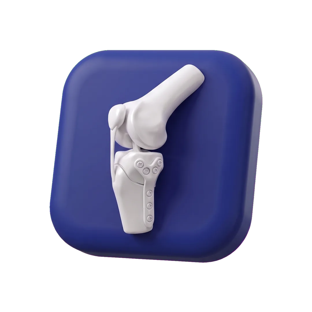
TPLO — treatment of a ruptured cruciate ligament
Surgery to stabilise the knee after a rupture of the cranial cruciate ligament in a dog. Planned using X-rays and performed under general anaesthesia with full monitoring, with a clear recovery plan.
What it involves
TPLO (tibial plateau levelling osteotomy) is an operation that does not “stitch” a ruptured ligament back together, but changes the geometry of the stifle joint so that the ligament is no longer needed. We make a controlled cut in the upper part of the tibia, rotate the fragment by a planned angle and stabilise it with a plate and screws. As a result, the femur no longer slides backwards off the tibia when the limb bears weight.
This is currently the best-documented method of treating cruciate ligament rupture in medium and large dogs. It allows early weight-bearing on the limb and usually gives better long-term function than techniques based on external sutures. It does not reverse existing degenerative changes, but it slows their progression.
During the operation we also assess the menisci — damage to them accompanies a ruptured ligament in a large proportion of cases and, if present, needs to be treated during the same procedure.
When it is used
We recommend TPLO once a rupture (complete or partial) of the cranial cruciate ligament has been confirmed in a dog, especially one over 15 kg, or an active or working dog. In smaller dogs other surgical techniques are also possible — we decide individually after the examination and X-rays.
The procedure should be carried out within a few days of the ligament rupturing, to avoid loading and damaging the menisci and overloading the other limb. We do not treat a ruptured ligament conservatively.
Indications: symptoms that should prompt a visit
- A dog suddenly started limping on a hind leg during play and has not put full weight on it since.
- Lameness of a hind limb eases with rest but returns after every longer walk.
- A dog sits with one hind leg stuck out to the side because it cannot bend the knee.
- The knee is thickened and the thigh muscles are visibly smaller than on the other side.
- A click can be heard in the knee during movement — possible meniscal damage.
- The veterinarian found a positive cranial drawer test or tibial compression test.
Conditions for which we use it in particular
Diagnostics beforehand
We make the diagnosis on the basis of an orthopaedic examination (knee stability tests) and digital X-rays of the knee in two projections, taken under sedation. On the lateral image we measure the tibial plateau angle — this determines the size of the correction and the choice of plate. The images therefore serve not only to confirm the diagnosis but to plan the operation.
Before the procedure the patient undergoes pre-anaesthetic assessment in line with standard STD-01: a clinical examination, a full blood count and biochemistry, and ASA classification. For older dogs or those with a heart murmur, a cardiological examination as well.
How it proceeds
The dog arrives in the morning, fasted. After premedication and pain relief (a multimodal regimen, STD-04, including a regional nerve block of the limb) we induce general anaesthesia. Throughout the procedure the anaesthetist monitors the ECG, oxygen saturation, capnography, blood pressure and temperature; the patient lies on a heated surface. We prepare the surgical field in line with our asepsis standard STD-05, and before the operation begins the team goes through the procedure checklist STD-03.
The procedure itself takes roughly 60–90 minutes. We open the joint, assess the meniscus and treat it if needed, perform the osteotomy, rotate the bone fragment by the planned angle and fix it with a plate. After closing the wound we take a check X-ray to confirm the position of the implant.
The dog wakes up in the in-patient hospital under the care of staff, with continuous pain control. It usually stays overnight; the extent of overnight supervision is determined by the lead veterinarian. It most often goes home the next day, walking on three or four legs.
Aftercare
The first 6–8 weeks involve strict restriction of activity: short walks on a lead only for toilet needs, no stairs, jumping or slippery floors; a collar until the stitches are removed after 10–14 days. We give pain relief and anti-inflammatory medication according to the written schedule. Clinical check-up after 2 weeks, check X-ray after 6–8 weeks — walks can be lengthened gradually only once bone healing has been confirmed.
Call us immediately if the dog suddenly stops putting weight on the leg, the wound starts to weep or the limb swells. A full return to activity usually takes 4–6 months.
Frequently asked questions
Can my dog walk after TPLO?
Yes, most dogs put weight on the limb within a few days. Walking is encouraged, but only on a lead and in short bouts, until an X-ray confirms that the bone has healed.
Does the plate stay in for good?
Usually yes. We remove it only if it causes problems (for example inflammation), which rarely happens.
Will the ligament in the other leg rupture too?
In some dogs, yes — the ligament ruptures on a background of degeneration, not only after an injury. Keeping body weight under control and regular check-ups allow this to be caught early.
Do you offer rehabilitation after the procedure?
We do not provide physiotherapy on site. You will receive a written plan of home exercises and, if needed, we will point you to a rehabilitation centre.
We manage every case individually. The scope, indications and course described above are indicative; we agree the exact diagnostic and treatment plan during the consultation. This content is for information only and does not replace a veterinary consultation.
Have questions about TPLO?
Call us or book a consultation. We will set out the diagnostic and treatment plan in writing, before you decide.