
Veterinary endoscope
A flexible video endoscope for examining the digestive and respiratory tracts, and a set of instruments for taking biopsies and removing foreign bodies.
What an endoscopy tower consists of
Click or hover over a part. The descriptions appear one after another.
- 1Monitor
A high-resolution image from the camera. On it the veterinarian assesses the mucous membrane and guides the instruments.
- 4Instrument table
Biopsy forceps, snares and graspers: instruments passed through the working channel to take tissue samples or remove a foreign body.
- 5Light source
Illuminates the organ being examined through a fibre-optic bundle running inside the scope.
- 7Wheeled base
The whole tower moves between the endoscopy room and the operating suite.
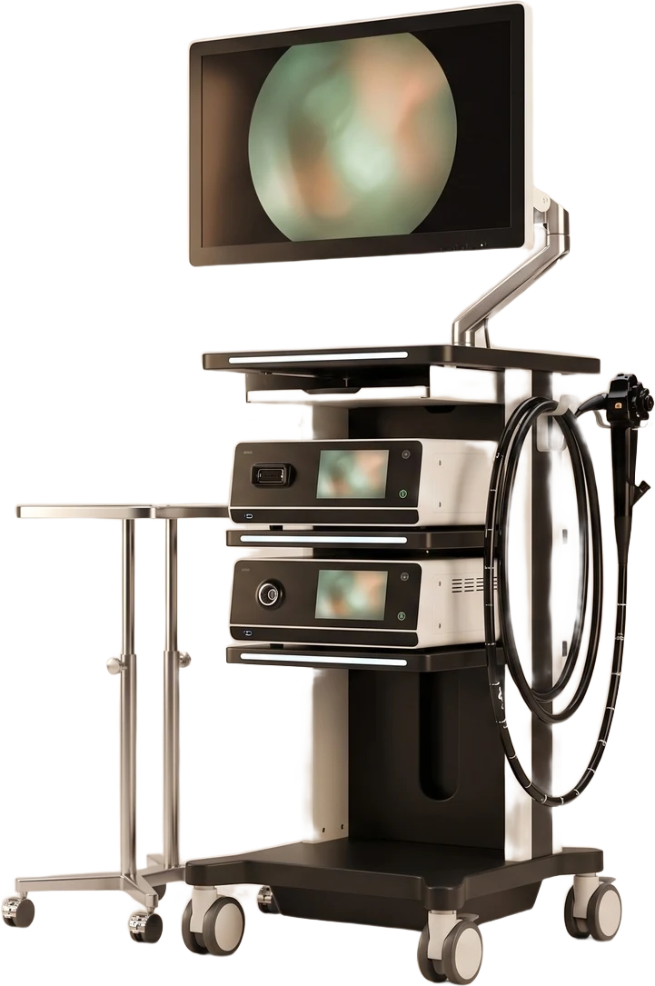
- 2Control handle
The dials bend the tip of the endoscope in four directions; this is also where instruments are inserted into the working channel.
- 3Image processor
Processes the signal from the camera at the tip of the scope. It saves images and video, which go into the patient's medical records.
- 6Flexible endoscope
A flexible scope with a camera and a working channel, inserted through the body's natural openings under general anaesthesia.
Illustrative diagram of an endoscopy tower. The arrangement of parts corresponds to our set-up, but the appearance of the devices may differ.
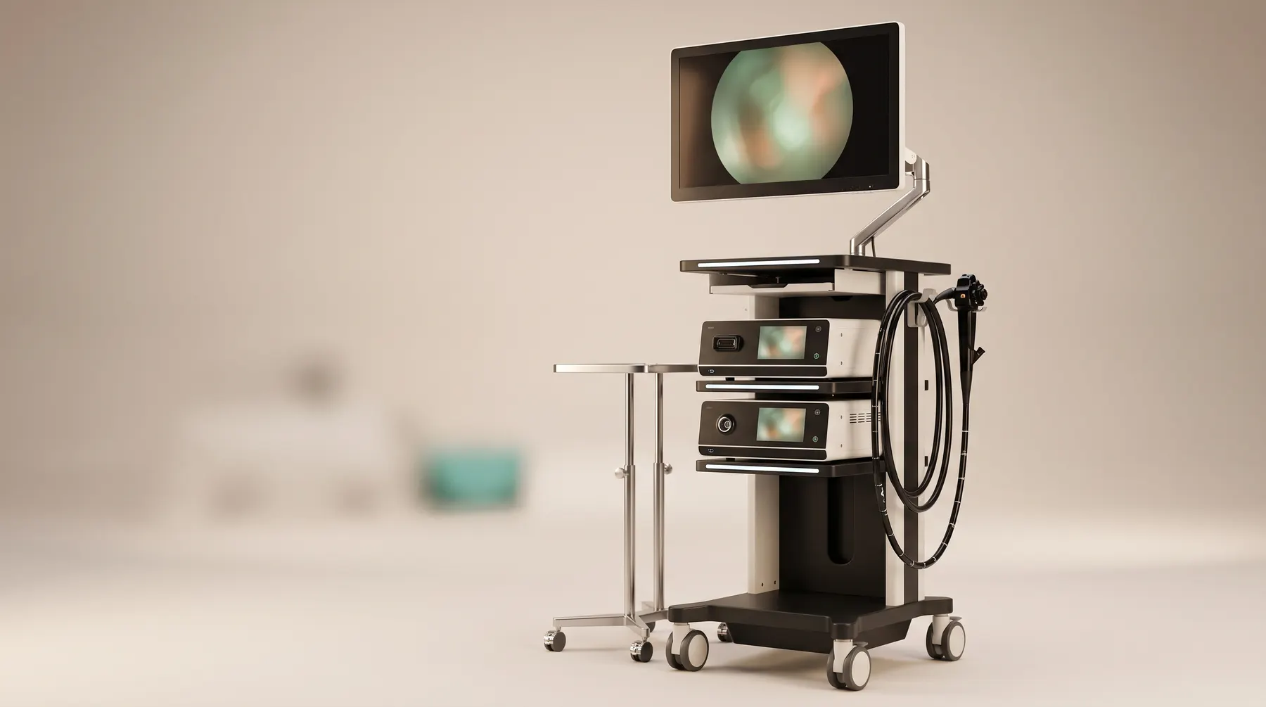 Endoscopy tower: complete set
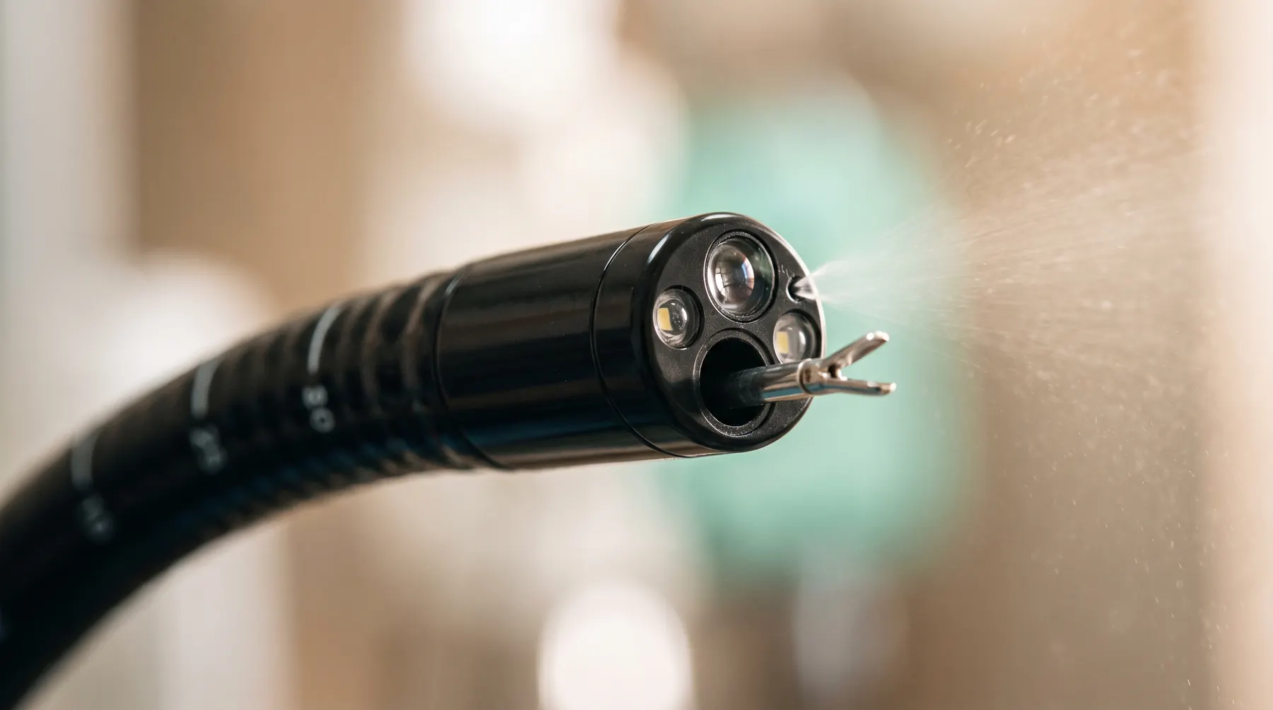 Tip of the endoscope: working channel, light, water irrigation
What it is and how it works
An endoscope is a flexible scope with a camera and a working channel, inserted through the body's natural openings under general anaesthesia. The image is shown on a high-resolution monitor; biopsy forceps, snares and graspers are passed through the working channel. This makes it possible to assess the oesophagus, stomach, duodenum, large intestine, trachea and bronchi, and the nasal cavity.
What it offers
Many foreign bodies (bones, toys, stones) can be removed without opening the abdomen. The patient goes home the same day or the next, with no surgical wound.
Endoscopic biopsy of the mucous membrane is the basis for diagnosing chronic intestinal disease (IBD), stomach tumours or chronic bronchitis, where ultrasound and blood tests give only a suspicion.