

Home/Specialties/Oncology
Specialty

Oncology

Diagnosis, staging and treatment of tumours — with a clearly explained prognosis and a plan the owner understands.

7 examinations and procedures6 items of equipment
Scope of this specialty

Examinations, procedures and services

We manage every case individually — the scope, indications and course described above are indicative; the exact diagnostic and treatment plan is agreed during the consultation.

Oncology · consultation

Oncology consultation

The first step when a dog or cat has a lump, an enlarged lymph node or a worrying test result. Assessment of the lesion, cytology usually on the same day, and a clear plan for the next steps.

DogCat
Oncology · examination

Fine-needle aspiration and tumour cytology

Sampling a tumour with a fine needle and examining the collected cells under a microscope — the quickest way to find out what a lesion is. Usually without sedation, often with a result on the same day.

DogCat
Oncology · examination

Biopsy and histopathology

Taking a piece of the lesion or all of it and assessment of the tissue by a pathologist — the definitive diagnosis of the type of tumour, its grade and whether the margins are clear after surgery.

DogCat
Oncology · examination

Tumour staging

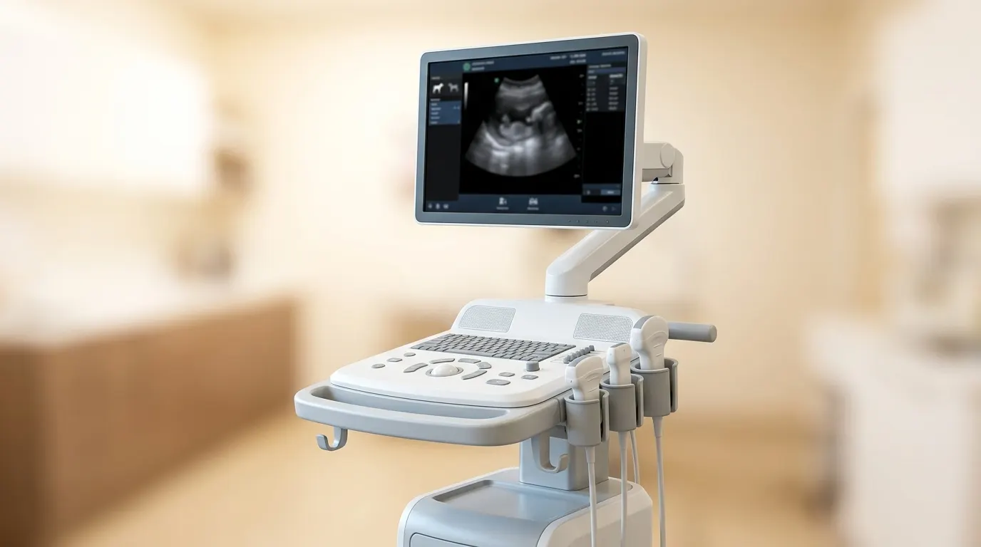
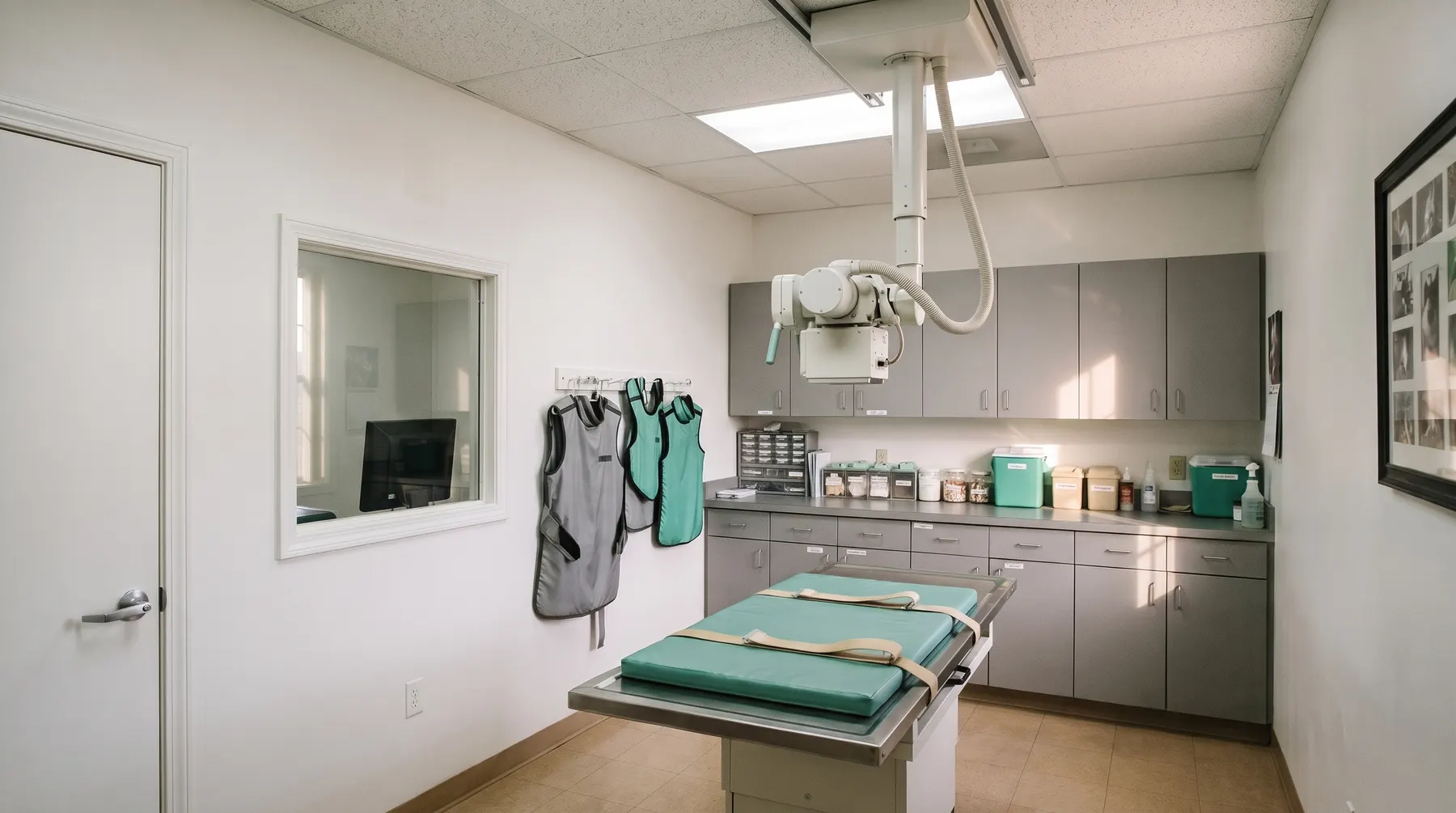
Checking whether and how far the tumour has spread: chest X-ray, abdominal ultrasound, lymph nodes, blood tests.

DogCat
Oncology · procedure

Surgical oncology — removing tumours with a margin

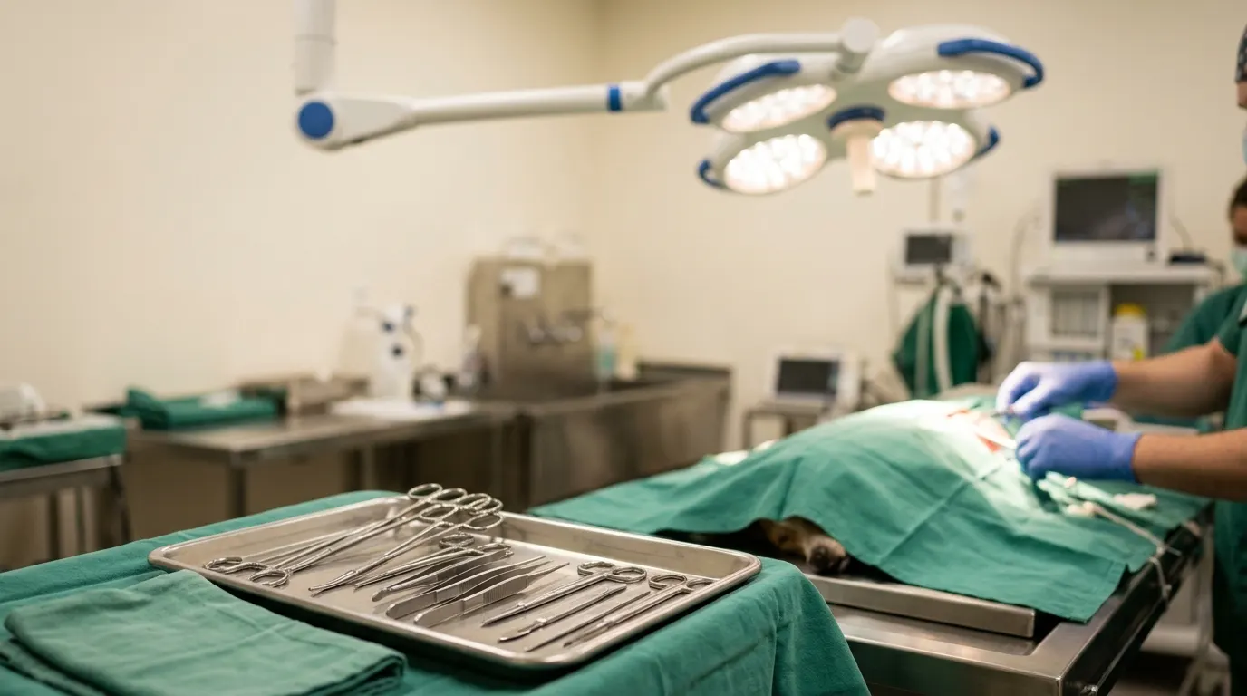
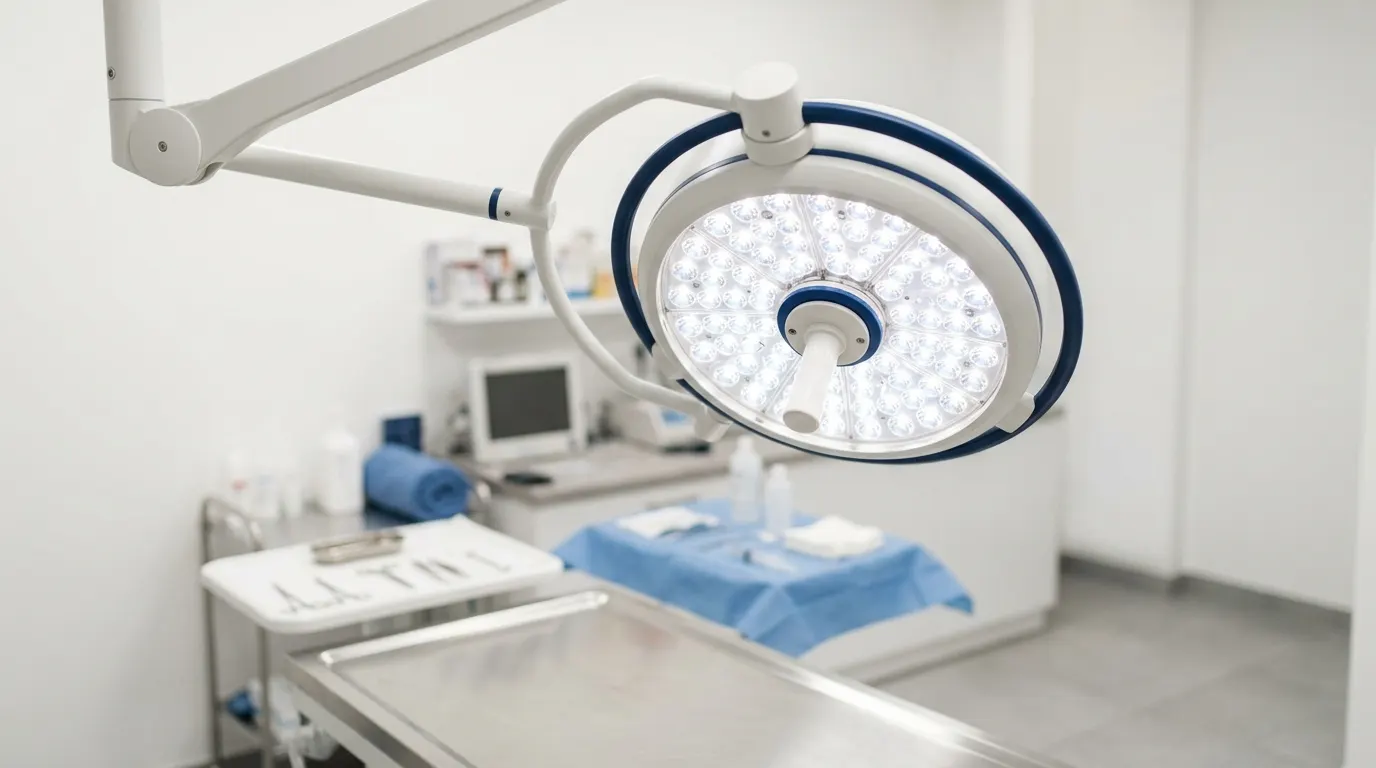
Surgical removal of a tumour planned on the basis of the diagnosis and staging — with a margin chosen to suit the type of tumour, in two operating theatres with full monitoring and histopathological assessment of every lesion.

DogCat
Oncology · treatment

Chemotherapy and drug treatment for cancer

Chemotherapy, targeted drugs and supportive treatment for cancer, given here on site. Where needed, we coordinate treatment with an external centre.

DogCat
Oncology · treatment

Mast cell tumour (mastocytoma) in a dog

Comprehensive care of a dog with a mast cell tumour — from cytology and staging, through surgery with an appropriate margin, to interpretation of the grade and a follow-up plan. The most common skin tumour in dogs.

Dog
Conditions and symptoms within our scope

What patients come to us with

Mast cell tumourthe most common skin tumour in dogs, variable in appearance and behaviour.
Lymphomaa cancer of the lymphatic system; in dogs usually with enlarged lymph nodes, in cats often the intestinal form.
Mammary gland tumoursin unspayed bitches and female cats; in cats mostly malignant.
Tumours of the spleen and liverfrom benign haematomas to haemangiosarcoma; risk of rupture.
Soft tissue sarcomasfirm, poorly movable lumps under the skin that infiltrate locally.
Oral tumoursmelanoma, squamous cell carcinoma, fibrosarcoma; early detection changes the treatment options.
Bone tumoursin large-breed dogs, with worsening lameness and swelling.
Lipomaa benign fatty tumour; cytology can confirm it without surgery.
Mast cell tumourmast cells have characteristic granules that are clearly visible on cytology.
Lymphomacytology of a lymph node is often enough to make the diagnosis.
Histiocytomaa benign tumour of young dogs that usually disappears on its own; cytology helps avoid unnecessary surgery.
Abscess and granulomainflammatory lesions that mimic a tumour and need completely different treatment.
Lead veterinarian for the specialty

Who manages the patients’ care

Profile photo
plain background
Lead veterinarian · oncology, dermatology, laboratory

Mateusz Podgórski, DVM

He leads diagnosis and treatment of oncology patients, from cytology and staging of the disease to a treatment plan agreed with the owner. Dermatology and laboratory diagnostics are his strong suit: he performs cytological examinations in the practice’s laboratory. He has a detective’s instinct: in his head he follows many threads at once, from laboratory results to the patient’s behaviour, and ties them together. He is a perceptive, experienced and dedicated doctor who can solve difficult diagnostic puzzles.

See the whole team
Frequently asked questions

Owners’ questions

Is every lump cancer?

No. Many lesions, especially lipomas and cysts, are benign. That is exactly why, instead of guessing, we sample the lesion with a needle and look at the cells.

Can taking a needle sample “spread” the tumour?

With a fine needle the risk is negligible and many times smaller than the risk of operating without a diagnosis. We know the exceptions (for example bladder tumours) and take them into account.

Wouldn't it be better to remove the whole lump straight away and send it for testing?

Sometimes, yes — with small skin lesions. With large or awkwardly placed tumours, an earlier diagnosis lets us plan the right margin and avoid a second operation.

Is staging necessary if we are planning surgery anyway?

Yes. Metastases in the lungs or lymph nodes change whether the operation makes sense at all, as well as its extent and aim. It is better to know this before the anaesthetic than after.

Why is the scar so large when the lump was small?

Because we remove not only the visible lump but also a margin of healthy tissue around and beneath it. A large scar is often the price of removing the tumour completely the first time.

Start with a conversation.

Reception will assess your enquiry and book an appointment with the right veterinarian.

This page is a translation of the Polish original. In case of doubt, the Polish version prevails. Polski