
Electrosurgery (electrocautery)
Precise cutting and coagulation of blood vessels with high-frequency current — less bleeding and a shorter procedure.
What an electrosurgical unit consists of
Click or hover over a part. The descriptions appear one after another.
- 2Active electrode
A handle with a tip with which the surgeon cuts tissue and seals blood vessels at the same time. It shortens the procedure and limits blood loss.
- 3Connection sockets
Connection of the active electrode and of the neutral electrode, which completes the circuit through the patient's body.
- 5Wheeled trolley
The unit stands on a trolley beside the operating table, outside the sterile field.
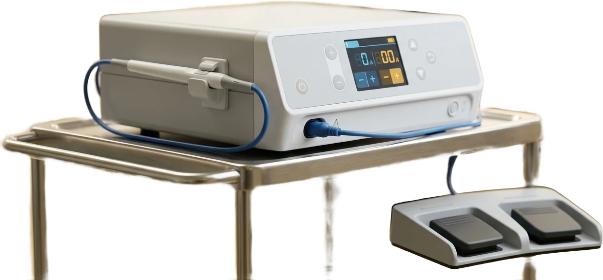
- 1Control panel
Selection of cutting and coagulation mode and of power; the display shows the current settings to the operator.
- 4Foot switch
Cutting or coagulation is activated with the foot, so that both of the surgeon's hands stay in the operating field.
Illustrative diagram of an electrosurgical unit. The appearance of the device may differ from ours.
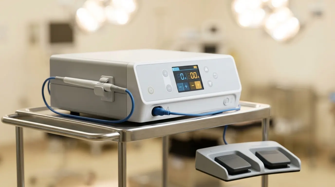 Electrosurgical unit
What it is and how it works
An electrosurgical unit makes it possible to cut tissue and seal blood vessels at the same time. It shortens the procedure, limits blood loss and improves visibility of the operating field, which is particularly important in splenectomy, mastectomy and the removal of well-vascularised tumours.
What it offers
Less blood loss means a shorter anaesthetic and faster recovery.
A clean operating field allows the surgeon to assess the margins of the lesion more precisely and to take material for histopathology without artefacts.